


Cervical osteochondrosis is a disease in which the spine and intervertebral discs are affected. Cervical osteochondrosis refers to deformed dorsopathies. Involutionary changes in the discs were observed as early as the age of 20 years. At the same time, they become more sensitive to loads, become less elastic, and lose lubricating fluid.
Most often, the pathology occurs in the elderly, but today the frequency in children and adolescents increases significantly. Neurologists are identifying cervical osteochondrosis using the latest diagnostic studies. After confirmation of the diagnosis, complex therapy is carried out with the most effective drugs, physiotherapy procedures and innovative methods of physical rehabilitation.
The name of the disease consists of two Greek terms "osteon" (bone) and "chondrosis" (cartilage). Cervical osteochondrosis begins with changes in the central part of the disc. The intervertebral disc loses moisture, decreases in size, this leads to convergence of the vertebral bodies and disruption of the nerve roots to the vessels. The spine receives nutrients from the surrounding tissues, which are harmful to the body. Compression of nerves and blood vessels causes spasm of the protective muscles, which causes pain as the disease progresses.

Which doctor treats this disease
Treatment of osteochondrosis is the field of activity of neurologists. However, in case of symptoms of neck osteochondrosis it is possible to consult a general practitioner. The neurologist selects the treatment medications for cervical osteochondrosis that have the least stress on the body, which is important for medication therapy.
To determine the presence of a pathological process in the cartilage tissue and cervical osteochondrosis, the patient is sent for a comprehensive examination. According to the results of the study, tactics have been developed on how to treat cervical osteochondrosis.
Interdisciplinary collaboration also allows for the treatment of comorbidities that a patient has. In addition, the patient receives complete informational assistance: treatment plan, extract on the cost of services, provision of information on specialist consultations and diagnostic measures.
ᲛReasons
Osteochondrosis of the cervix develops under the influence of various provocative factors. Certain causes of cervical osteochondrosis have not been identified. The disease is often associated with metabolic disorders and aging of the spine.
Researchers suggest that cervical osteochondrosis develops for the following reasons:
- Excessive stress on the spine. High load on the spine is observed during incorrect shoes, flat feet, obesity, prolonged sitting position;
- Metabolic disorders. Deficiency of vitamins, minerals, disorders of calcium metabolism can be the causes of degenerative processes in the spine;
- Congenital and acquired anomalies of the spine and yoga apparatus (thickening of ligaments, lumbarization, sacralization);
- Pathologies of the gastrointestinal tract, leading to insufficient absorption of nutrients;
- Infection, intoxication;
- Injuries, bruises, fractures of the spine, resulting in disruption of the blood supply and innervation of the spinal column, leading to their dystrophic disorders;
- Ს comma;
- Wearing shoes with heels;
- Pregnancy, especially multiple pregnancies;
- Autoimmune damage to connective tissue, abnormal structure of collagen types 1 and 2;
- Occupational hazards (lifting heavy loads, prolonged vibration, working in a sitting position with constant head tilt);
- Atherosclerotic and other changes in the vertebral arteries;
- Spinal curvature (kyphosis, scoliosis, kyphoscoliosis).
An important risk factor for the development of cervical osteochondrosis is a loaded inheritance. This fact confirms the presence of osteochondrosis in children when the spine is not yet overloaded.
Degrees
Due to the special structure of the spine, it can perform its functions. The main structural unit is the spinal movement segment (VMS). It consists of two adjacent vertebrae, the intervertebral disc and the musculoskeletal system. Osteochondrosis causes dystrophic-degenerative processes, first in the intervertebral disc and then in the spine. By defeating one spine, its functions are provided to adjacent individuals. This leads to an increase in the load of the loaded segment and loss of mobility.
During the development of cervical osteochondrosis, doctors distinguish several stages:
- First degree of cervical osteochondrosis. Because the intervertebral disc is deprived of its own blood supply and receives nutrients from the surrounding tissues, it is subject to degenerative changes. Osteochondrosis in stage 1 development is characterized by destruction of the nuclear pulp and fissures in the annulus fibrosis. Clinically it is manifested by acute or persistent local pain in the neck (cervicalgia) and stiffness;
- Second degree osteochondrosis of the cervical spine. At this stage the destruction of anol fibrosis continues, abnormal mobility and instability of the malleolus occurs. Patients complain of neck pain, aggravated by physical exertion, tilting of the head, or in a certain position;
- The third stage of the disease is characterized by complete destruction of the anal fibrosis. The gelatin nucleus is not fixed. Herniated discs can be detected and cause severe pain. At this point, due to poor fixation of the SMS, a curvature of the spine may occur;
- In the fourth stage of the disease, the intervertebral disc is replaced by connective tissue, acting on other adjacent segments. Develops spondyloarthritis, arachnoiditis. The joints become completely immobile - ankylosis develops. Bone tissue grows around the affected area - osteone is formed. In the fourth degree of cervical osteochondrosis there are strong symptoms: severe pain radiating to the arm, bump, area between the shoulder blades, sensitivity disorders.

Symptoms and signs
At the initial stage, the signs of cervical osteochondrosis may be nonspecific: dizziness, headache, weakness, cramps during movement. As the disease progresses, the following symptoms develop:
- Severe pain in the neck and shoulders;
- Hand numbness;
- Dizziness;
- Increased blood pressure;
- Movement coordination disorder;
- Increased sweating.
There are several syndromes that arise with the development of abnormal conditions of the muscles of the spine and cervical spine:
- Cervical migraine syndrome.
- Spinal artery syndrome.
- Hypertensive syndrome.
- Heart syndrome.
- Radical syndrome.
They occur when the nerve endings are damaged, the arteries and veins become tense during the development of the disease. The most dangerous complication is considered to be vertebral artery syndrome. Disrupted blood flow to the arteries that feed the brain and spinal cord. The patient's hearing decreases, vision decreases, constant dizziness develops. The patient may lose consciousness while driving due to a severe disturbance of blood flow.
Compression of the nerves responsible for innervating the muscles of the chest and diaphragm results in pain in the heart that is not related to heart disease but at the same time can cause tachycardia, arrhythmia, and hypotension. Intracranial pressure increases, nausea, vomiting, and severe headache manifest themselves due to deterioration of blood flow from the brain.
As a result of neck strain develops radicular syndrome - severe pain occurs in the neck, shoulders, shoulder blades and back of the head. With this syndrome, the arms and neck area swell. With cervical migraine syndrome, the patient suffers from severe occipital pain, often accompanied by nausea and vomiting.
Reflex syndromes occur when the spinal roots are not yet damaged. Patients complain of pain in the neck, head (especially the back of the head), arms on one or both sides. Reflex pain, unlike radicular pain, is not combined with sensitivity disorders. Cervical pain can be dull, painful. Acute "lumbago" pain is called cervical pain. Spasm and muscle pain, pain in the intervertebral discs are noted. Signs of cervical osteochondrosis are exacerbated by discomfort, head tilting, coughing, and physical exertion. Signs of epicondylosis, humeroscopic periarthrosis, and hand-shoulder syndrome occur due to nerve impulses from the fibrosis of the affected segment anus, leading to compensatory muscle spasm.
Radical syndromes are accompanied by motor activity and sensitivity. At the same time, the nerves, blood vessels are impaired, venous and lymphatic outflow in the pathological focus is disrupted as a result of narrowing of the intervertebral canal. The pain of radicular syndrome is acute, intense. A common cause of spinal nerve entrapment is the formation of a hernia. Muscle tone decreases in the area of abnormal focus. With radiculosemia, in addition to the nerves, the vessels are compressed.
If the phrenic nerve is involved in the prenatal process, heart syndrome occurs. It manifests itself as burning, sharp pain in the left side of the chest with radiation to the interduloid region. The name of the syndrome is due to the fact that the nature of the pain is similar to that of an angina attack. The main difference between angina pain is that it is relieved after taking nitroglycerin, it can occur in the rest of the condition and is combined with cardiac arrhythmias (tachycardia, arrhythmia).
Signs of cervical osteochondrosis depend on the localization of the pathological process. With a cervical spine injury, the blood supply to the brain is disrupted due to compression of the cerebral arteries. This causes headaches (especially in the occipital area), dizziness, loss of consciousness, high blood pressure. Dizziness in cervical osteochondrosis is caused by a decrease in blood flow to the inner ear. Patients also suffer from nausea, vestibular and ocular symptoms.
With combined damage to the spine, they talk about cervical osteochondrosis. The disease is manifested by the following symptoms:
- Dizziness;
- Pain in neck and arm;
- Tingling, creeping sensation in the upper limb;
- Intercostal neuralgia.
Diagnosis
Osteochondrosis of the cervix is a chronic disease that can lead to the formation of hernias and compression of the spinal cord. It is therefore important to make an accurate diagnosis in a timely manner and start therapy. The following types of instrumental diagnostics are used to diagnose cervical osteochondrosis:
- Spinal spondylography or X-ray. This method of research is painless, informative and does not require special training. X-ray of the spine allows you to assess its anatomical and functional characteristics. The picture focuses on the structure of the spine, their relationship to each other, including the distance, the lumen of the spinal canal;
- Computed tomography - mainly provides information about the condition of bone tissue, allows you to determine the narrowing of the spinal canal and herniated disc;
- Magnetic resonance imaging - allows you to detect soft tissue changes. The MRI image clearly shows changes in the intervertebral discs and spinal cord.

Medication treatment
Treatment of cervical osteochondrosis consists of medication and non-drug therapy. Even after complete cure, neurologists take preventive measures to rule out recurrences of the disease. For the treatment of osteochondrosis of the cervix during the acute period, doctors prescribe drugs to patients of the following pharmacological groups:
- Non-narcotic analgesics. They are taken orally or injected intramuscularly to achieve a rapid effect;
- Nonsteroidal anti-inflammatory drugs;
- B group vitamins in large doses.
Diuretics are used to reduce fluid retention in the spinal cord and surrounding tissues. Antihistamines enhance the action of analgesics. Muscle spasms are eliminated by muscle relaxants. With prolonged acute pain syndrome, neurologists block the nerve.
Chondroprotectors are used to improve metabolic processes in the intervertebral disc. These drugs increase the content of glycosaminoglycans, increase the density of the intervertebral discs, elasticity and shock absorption.
Dizziness pills
Patients often have dizziness during cervical osteochondrosis. To reduce them, doctors prescribe nonsteroidal anti-inflammatory drugs. Non-steroidal anti-inflammatory drugs belonging to different groups differ in mechanism of action and effect, therefore, only a qualified specialist can prescribe the appropriate drug.
It is important to remember that cervical osteochondrosis medications can not be taken without a doctor’s appointment. Nonsteroidal anti-inflammatory drugs have side effects, so before prescribing them, the neurologist will determine the presence of contraindications in the patient and the required dose. Anti-vertigo drugs for cervical osteochondrosis can improve a patient’s quality of life.
Injections for osteochondrosis
Injections of cervical spine osteochondrosis contribute to pain during exacerbation. With this method of taking the drug, the effect occurs quickly. Neurologists use a variety of injections.
Nurses take drug solutions subcutaneously, intramuscularly, or intravenously. During the period of exacerbation of the disease, only injectable drugs for cervical osteochondrosis have only a symptomatic effect.
Headache treatment
Headache is a symptom found in a variety of disorders. However, cervical osteochondrosis is characterized by severe headaches. Head movements increase symptoms, so doctors prescribe painkillers and nonsteroidal anti-inflammatory drugs to eliminate it.
Non-drug therapy methods
Complex non-drug therapy for cervical osteochondrosis includes:
- Protective mode - when the roots are dug, patients are lying on a solid surface;
- Მ massage;
- Physiotherapy exercises;
- Spine traction;
- Physiotherapy procedures.
For cervical osteochondrosis, massage is used to reduce pain and swelling, improve peripheral blood supply, and relieve muscle spasm. A contraindication to this procedure is the presence of severe pain. Massage in the direction of lymph drainage to the neck and back. Particular attention is paid to the intercapsular and paravertebral zones.
Therapeutic gymnastics for cervical spine osteochondrosis aims to eliminate muscle spasm and strengthen the muscle frame. Since spinal instability often occurs in the cervical spine, the exercise therapy instructor conducts individual lessons during which he or she teaches the patient to perform the exercises safely. Some authors recommend conducting physical therapy classes in the shunt collar.
To improve the mobility of the cervical spine, rehabilitation therapists recommend the following exercises:
- Neck bend and extension. Bend your head forward towards your waist, without stretching your shoulders forward, then backwards. Hold the incline for 3 seconds, repeat each exercise 8-10 times;
- Neck bend. Rotate the neck first to the left until it stops, then to the right, without changing the position of the shoulders and the level of the chin;
- Lower your head until it stops. Then bend your head back, without changing shoulder level. Hold the position for 5 seconds.
The following exercises have been developed to strengthen the neck muscles:
- Place your hand behind your head. Lean your head back, lean on your hand;
- Place your hand temporarily in the region. Resist while tilting your head;
- Put your hand on your forehead, resist it, lean your head forward;
- Lean your head to the right and back with your left hand. Repeat the exercise on the other hand.
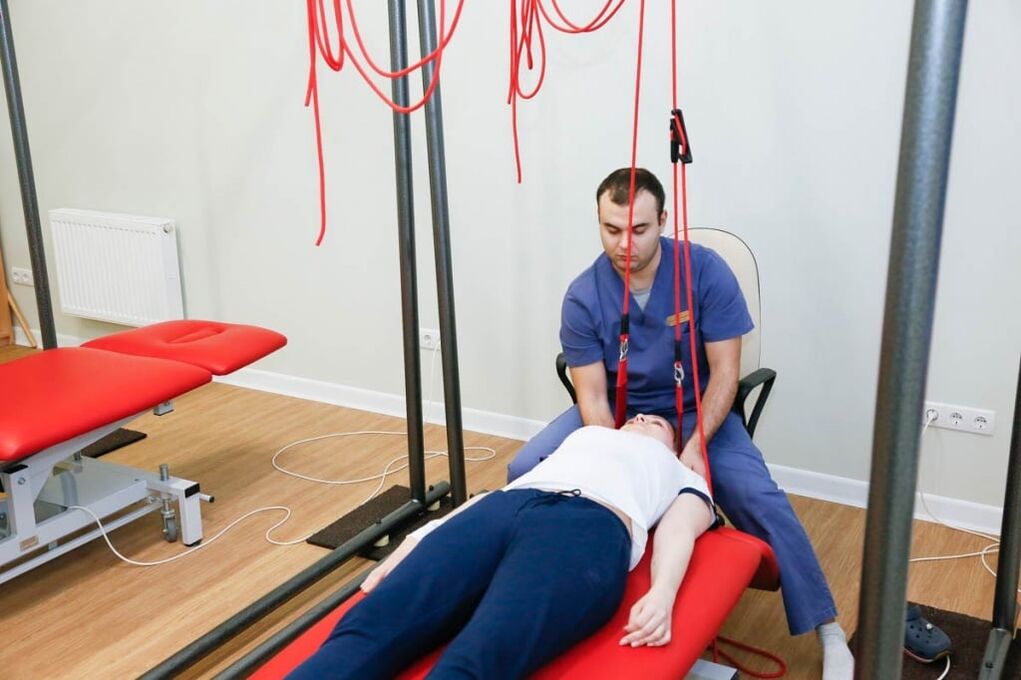
Autorevitation therapy is the exact name of the spine attraction procedure. It is carried out using special devices. The goal of therapy is to reduce muscle spasm and restore the correct position of the spine. Spinal traction is performed by a doctor to prevent complications.
The following physiotherapy procedures are used to improve blood supply, relieve swelling, and relieve pain in the pathological focus:
- Diadynamic currents. During this procedure, using a special device, low-frequency currents are used, which stimulate the muscles, relieve spasms and pain. Has a positive effect, improves tissue trophism;
- Ultraviolet radiation. Under the influence of ultraviolet radiation, the metabolism of vitamin D is improved, the calcium content is increased, bone tissue is strengthened;
- Ultrasound effect - used to accelerate blood flow, spasmolytic and reparative action. Ultrasound can penetrate deep into tissues, sometimes it is used for better absorption of healing substances;
- Amplipulse therapy - allows you to relieve pain by blocking nerve impulses from painful focus.
In the acute period of the disease, which lasts 4-7 days, antispasmodics, irritants are used to reduce pain. The patient is provided with peace. Cervical spine immobilization is performed using a shunt collar. Exercise therapy and massage are contraindicated. Use ultraviolet radiation.
The duration of the subacute period is 29 days. After complete recovery the patient should rest for a few days. Then you can start a course of rehabilitation therapy. In the chronic course of the disease the patient is prescribed muscle relaxants, chondroprotectors, B vitamins, in pain - analgesics, nonsteroidal anti-inflammatory drugs. Physiotherapy exercises, massage are provided. The patient is relieved by physiotherapy procedures (amplitude, alternating current exposure), spinal traction occurs.

Food
Proper nutrition of osteochondrosis is an important condition for achieving remission. Progression of cervical osteochondrosis will stop with diet and treatment. Neurologists know how to treat cervical osteochondrosis, so they create a set of therapeutic measures, including procedures, exercise therapy, proper nutrition, and lifestyle changes.
Many patients ask neurologists how to treat cervical osteochondrosis and to what extent is food restriction limited. Specialists create individual nutrition programs that take into account the patient's preferences. The diet for osteochondrosis is based on a balanced, low-fat diet rich in nutrients. The patient's daily diet contains calcium-containing foods.
How to sleep with cervical osteochondrosis
For patients with diseases of the musculoskeletal system, the question of how to sleep properly with cervical osteochondrosis is relevant. Sleeping on your stomach causes further development of the disease, so it is best to avoid sleeping in this condition. The most optimal positions are at the back.
Cervical osteochondrosis progresses when lying on a soft mattress in bed. Therefore, experts recommend giving preference to elastic mattresses, as well as moderately soft cushions. If a patient is asked about cervical osteochondrosis, experienced specialists will tell you which beds are safe for sleep.
Prevention
To prevent the onset or progression of cervical osteochondrosis, doctors recommend:
- Maintain the correct posture;
- Live an active lifestyle, take a break from work;
- Do regular physiotherapy exercises;
- Sleep on solid and level surfaces, orthopedic mattress and pillow;
- Avoid bad habits, especially smoking;
- Choose shoes taking into account the physiological structure of the foot;
- Do not carry the bags on one side, this will cause the spine to bend;
- Live a healthy lifestyle, eat right, eat lots of fruits and vegetables;
- Long does not sit with his head tilted;
- Walk on the . urva.
To improve blood circulation, massage therapy should be performed regularly.